My Pathology Report
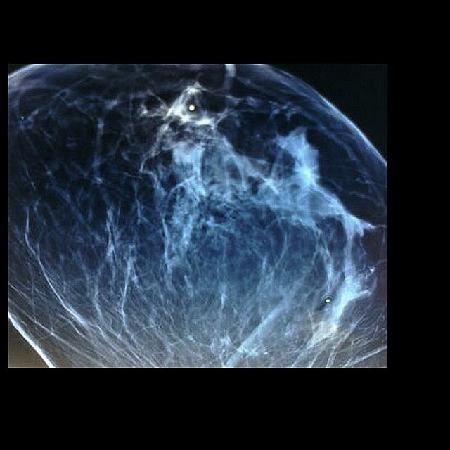


The pic above is one from my actual mammograms-pretty interesting, I think-you can compare it to the one below, which isn't mine-if you look closely you'll see a solid BB dot which they put on your breast to basicly hone in on what they want to see.
FINAL PATHOLOGIC DIAGNOSIS:
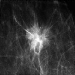
DCIS in someone else's boob
Ductal carcinoma in-situ (DCIS); Intermediate and high nuclear grade; solid, cribriform and micropapillary type.
DCIS admixed with fibrocystic change, total combined area approximately 4.0cm diameter
.>DCIS focally less than 0.1cm from cauterized surgical margin.
No invasive carcinoma identified.
Microcalcifications associated with ductal carcinoma in-situ and proliferative breast disease.
Fibrocystic change.
Estrogen receptor; Positive, 50% of nuclei staining with moderate intensity (2+)
Progesterone receptor; Positive, 90% of nuclei staining with strong intensity (3+)
